
Mastering Careers & Skills
Diagnostic Medical Sonography Exams (ARDMS) · free sample from the book itself
Diagnostic Medical Sonography Exams (ARDMS) — Exam Prep Series — 2026 Edition. Every rationale explains why the key is right and why each wrong option is wrong.
Q1 — Key: B. Tag: CRAFT. The sonographer conducts an examination — reviewing the indication, acquiring images and measurements systematically, and presenting a study that answers the clinical question (§1.1). A reduces the work to speed, which §1.1 rejects: a fast study that misses the question has failed. C assigns the final diagnosis to the sonographer, which belongs to the interpreting physician (§1.5). D discards the indication, when the indication is what the whole examination is organized to answer.
Q2 — Key: C. Tag: CRAFT. Sycamore Street is this book's fictional specimen service, labeled as fictional at first use, whose stated facts are its own and no one else's (§1.1). A claims a real department and reprinted records; no real records appear anywhere in the book. B assigns the service to ARDMS, which certifies individuals and runs no service in this book. D invents a size ranking no source states.
Q3 — Key: A. Tag: CRAFT. The canon is 5 scan rooms, 7 credentialed sonographers and a lead sonographer, 2 students on clinical rotation, and 38 scheduled examinations in a day across abdomen, obstetric, gynecologic, and vascular lists (§§1.1–1.2). B understates both rooms and volume. C denies the students the book explicitly places on rotation. D contradicts the team structure the whole chapter describes.
Q4 — Key: D. Tag: ABDO. Mrs. Delgado's canon normal study carries a common bile duct of 4 mm and a liver span of 14.6 cm (§1.2). A substitutes a duct value far outside the canon normal study. B substitutes a liver span the canon does not give. C is false because the canon study is defined precisely by its stated measurements — establishing normality completely requires them.
Q5 — Key: B. Tag: ABDO. A fed gallbladder has contracted, so it cannot be assessed the same way as a fasted one; §1.2 teaches preparation as part of the examination itself, not as paperwork. A calls fasting a clerical preference, denying the physiologic reason the instruction exists. C invents a prohibition on transducer use after meals. D invents a Doppler prohibition that no part of the chapter states.
Q6 — Key: A. Tag: VASC. Mr. Okafor's canon values are right internal carotid PSV 246 cm/s, EDV 104 cm/s, ICA/CCA ratio 4.1, and right ABI 0.72 (§1.2). B drops the carotid values that define his vascular-list case. C gives Ms. Nguyen's obstetric measurement to the wrong patient. D gives Mr. Petrov's aortic value to the wrong patient — the canon assigns each value set to one patient so chapters stay consistent.
Q7 — Key: C. Tag: OBGY. Ms. Nguyen's first-trimester canon values are a crown-rump length of 61 mm and a nuchal translucency of 1.8 mm (§1.2). A names only her later-pregnancy fluid value and misses the first-trimester pair the stem asks for. B gives Mr. Petrov's renal index to the wrong patient. D halves her canon biophysical profile of 8 out of 8.
Q8 — Key: D. Tag: ROLE. The interpreting physician reads the completed study, correlates it with the clinical history and other imaging, and issues the final diagnostic report (§1.5). A assigns examination conduct to the physician, when the sonographer conducts the study. B names a scheduling task belonging to service operations and the lead. C names an educational task belonging to the program and its supervisors, not the interpreting role as defined.
Q9 — Key: C. Tag: ROLE. A limitation is annotated honestly so the interpreting physician can weigh the study's limits rather than guess at them (§1.5). A conceals the limitation, leaving the physician to trust a study whose completeness is unknown. B assumes repetition removes limits such as bowel gas or a position the patient cannot hold, which it often cannot. D claims final-diagnosis authority the sonographer does not hold.
Q10 — Key: B. Tag: CRAFT. Abdomen sonography examines the liver, biliary system, pancreas, urinary system, retroperitoneum and great vessels, and included superficial structures (§1.3). A assigns chamber work to vascular technology, when cardiac examination is echocardiography's territory. C reverses gynecology's subject, which is the female pelvis. D reverses obstetrics' subject, which is pregnancy.
Q11 — Key: D. Tag: CRAFT. The fenced set reports the 2025 median pay as $96,590 per year ($46.44 per hour) (§1.6). (Source: U.S. Bureau of Labor Statistics, Occupational Outlook Handbook, "Diagnostic Medical Sonographers.") A gives the lowest-decile boundary, $67,820, not the median. B gives the highest-decile boundary, $129,370, not the median. C misreads any median as a guaranteed starting wage, which §1.6 explicitly denies.
Q12 — Key: A. Tag: CRAFT. The fenced set reports 14 percent growth for 2025–35, described as much faster than average, an employment change of +12,900, and about 6,000 openings per year (§1.6). (Source: U.S. Bureau of Labor Statistics, Occupational Outlook Handbook, "Diagnostic Medical Sonographers.") B reverses growth into decline. C denies a projection the source plainly publishes. D converts a national projection into a personal hiring promise, which §1.6 states no figure makes.
Q13 — Key: C. Tag: CRAFT. In the fenced employer distribution, hospitals hold the largest share at 58 percent, followed by offices of physicians at 21 percent (§1.6). (Source: U.S. Bureau of Labor Statistics, Occupational Outlook Handbook, "Diagnostic Medical Sonographers.") A, B, and D each attach the 58 percent figure to a setting whose own stated share is 5, 21, and 2 percent respectively — the exact label-stripping error §1.6 fences against.
Q14 — Key: B. Tag: CRAFT. A median is the middle of everyone counted — half earn more, half earn less — across experience and region, and §1.6 states it is not a starting wage, an offer, or a promise. A misreads the median as an entry wage. C describes a maximum, not a middle value. D invents a wage-setting power for the registry, which certifies individuals and sets no wages.
Q15 — Key: D. Tag: ROLE. The book teaches knowledge and prepares for the examinations while conferring no credential, no eligibility, and no authorization to scan any person (§1.6). A claims credentialing and authorizing powers no book holds. B promises an examination outcome the book expressly never promises. C claims the book replaces an accredited program, when accredited programs and supervised clinical experience are the doors Chapter 2 teaches.
Every imaging profession has a subject — the thing it looks at and the way it looks. For diagnostic medical sonography, the subject is the living body examined with sound. A diagnostic medical sonographer uses a transducer that sends high-frequency sound into the body and receives the returning echoes, and a scanner that turns those echoes into images, waveforms, and measurements that a physician can interpret. The work is called sonography when the emphasis is on producing the study, and ultrasound when the emphasis is on the sound technology itself; in everyday practice the two words travel together, and this book uses sonographer for the person, sonography for the craft, and ultrasound for the sound and the examination it makes possible.
That short definition already carries the profession's central discipline. The sonographer does not simply take pictures. The sonographer conducts an examination: reviewing why the study was ordered, choosing how to approach the anatomy in question, acquiring images and measurements in a systematic order, recognizing when what appears does not fit what was expected, extending the examination within protocol when the findings require it, and presenting the completed study so that the interpreting physician can answer the clinical question that sent the patient to the scan room. A technically beautiful image that does not answer the question has failed, and an unglamorous image that answers it completely has succeeded. Learning to judge your own work by that standard — did the study answer the question, completely and honestly? — is the habit this entire book is built to form.
To keep the teaching concrete, this book carries one worked example through its chapters: the Sycamore Street Imaging Service, a fictional hospital-affiliated outpatient imaging service used in this book as a specimen — a worked example whose stated facts are its own and no one else's. Sycamore Street is not a real employer, and no real person works or is examined there. Its stated facts are these: 5 scan rooms, 7 credentialed sonographers and a lead sonographer, and 2 sonography students on clinical rotation; a day of 38 scheduled examinations across abdomen, obstetric, gynecologic, and vascular lists. Four fictional patients recur so that values stay consistent from chapter to chapter: Mrs. Delgado, 58, on the biliary and liver list; Mr. Okafor, 66, on the vascular list; Ms. Nguyen, 29, on the obstetric list; and Mr. Petrov, 71, on the aorta and renal list. When a number belongs to Sycamore Street or to one of these patients, the text says so. When a number belongs to the real world, it comes only from a named source in this book's research register, cited where it prints. Knowing which numbers are teaching examples and which are sourced facts is itself a professional habit, and this first chapter practices it from the start.
Ask what a sonographer actually does and the most honest answer is a day, described plainly. At Sycamore Street, the stated day holds 38 scheduled examinations across four lists, and the arithmetic of the service is worth noticing once: 38 examinations shared among 7 credentialed sonographers is a little over five studies per sonographer before add-on examinations, urgent requests, and the inevitable study that takes longer than its slot are counted. No sonographer works that day as an isolated pair of hands. The lead sonographer holds the list together — triaging urgent requests, rebalancing rooms when a study runs long, and serving as the first consult when a finding does not fit. The two students observe and participate only within their supervised clinical rotation, under the authority of the program and the service together, a boundary Chapter 2 examines in full.
Picture the shape of one sonographer's morning, in knowledge terms — what the work is and why it is sequenced as it is, not as a procedure to copy. The day begins before the first patient enters the room: reviewing the day's orders and the clinical indication for each — the symptom, finding, or question that justifies the study — checking prior imaging so the new study can be compared with what came before, and confirming that preparation requirements have been met. Preparation is not clerical detail. A biliary examination ordered to assess the gallbladder and ducts means something different in a patient who has fasted as instructed than in one who has just eaten, and the sonographer who knows why — a fed gallbladder has contracted, and a contracted gallbladder cannot be assessed the same way — understands the preparation as part of the examination itself. Mrs. Delgado's normal study on the biliary list, with a common bile duct measuring 4 mm and a liver span of 14.6 cm in this book's canon, is useful teaching precisely because it is normal: the sonographer had to know the anatomy, the protocol, and the measurements well enough to establish normality completely, not merely to fail to find disease.
The middle of the day is the examination itself, repeated with variation. Each study follows the same underlying pattern even though the anatomy and the questions differ entirely: confirm the patient and the order, explain the examination, position and scan systematically, measure according to protocol, interrogate with Doppler where the protocol and the findings call for it, and watch continuously for findings that change what the study must include. On the vascular list, Mr. Okafor's study carries canon values of a right internal carotid peak systolic velocity of 246 cm/s, an end-diastolic velocity of 104 cm/s, an internal carotid to common carotid ratio of 4.1, and a right ankle-brachial index of 0.72 — values the later vascular chapters teach you to reason with. On the obstetric list, Ms. Nguyen's canon values include a crown-rump length of 61 mm and a nuchal translucency of 1.8 mm in the first trimester, and, later in pregnancy, an amniotic fluid index of 13.5 cm with a biophysical profile of 8 out of 8. On the aorta and renal list, Mr. Petrov's canon values are an infrarenal aortic diameter of 4.8 cm and a right renal resistive index of 0.68. Hold those numbers lightly for now. Their purpose in this chapter is to show you what a sonographer's output actually is: not an impression, but images plus numbers, obtained by a defensible method, that another clinician will act on.
The day ends, as it began, with communication. Each completed study is documented, archived, and accompanied by the sonographer's preliminary communication in the form the service requires — the subject of Chapter 36 in full — and any finding that requires immediate action is communicated immediately, not filed and forgotten until a routine report appears. Threaded through the whole day is work a schedule never lists: cleaning and preparing rooms and transducers between patients under the infection-control discipline of Chapter 4, helping patients who move slowly or anxiously, coordinating with nursing, transport, and the front desk, and teaching — explaining to a patient what the examination involves, and explaining to a student why a measurement was taken where it was taken. At the point in the day where equipment is introduced, what the newcomer sees first is the machine at the center of the room.
Figure 1.1 (Class P) — A cart-based ultrasound system stands ready in a scan room, its display screen blank, with the transducer holders and control panel arranged for the next examination.
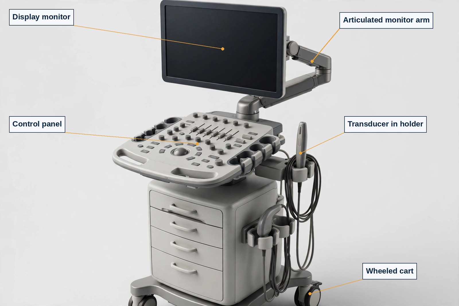
Figure 1.1 — A cart-based ultrasound system stands ready in a scan room, its display screen blank, with the transducer holders and control panel arranged for the next examination.
Sonography is one craft practiced in several territories, and a beginner needs the map whole before choosing where to stand on it. The national occupational account that Chapter 1 and Chapter 2 both draw on names the field's specialties inside its entry: the U.S. Bureau of Labor Statistics entry covers diagnostic medical sonographers with cardiac, musculoskeletal, pediatric, obstetric and gynecologic, and vascular specialties named in the entry. (Source: U.S. Bureau of Labor Statistics, Occupational Outlook Handbook, "Diagnostic Medical Sonographers.") This book teaches the territories that correspond to its four examinations — the physics examination common to all tracks, plus abdomen, obstetrics and gynecology, and vascular technology — and names the wider field honestly so that no reader mistakes this book's scope for the profession's whole extent.
Abdomen sonography examines the organs and vessels of the abdomen and the superficial structures the abdomen outline includes: the liver and portal system, the biliary system, the pancreas, spleen, and gastrointestinal tract, the urinary system, the retroperitoneum and great vessels, and structures from the thyroid to the scrotum and the body cavities. Its clinical questions are the everyday questions of internal medicine and surgery — pain, abnormal laboratory values, a palpable mass, suspected stones, obstruction, fluid, or infection — and its craft is systematic completeness: a survey that covers the territory in an order that leaves nothing unexamined merely because it was hard to see. Obstetric and gynecologic sonography joins two related practices. Gynecology examines the female pelvis — uterus, endometrium, ovaries, and adnexa — across the menstrual cycle and the lifespan, for pain, bleeding, masses, and the location of an intrauterine contraceptive device, among many other questions. Obstetrics examines pregnancy: confirming and dating it, surveying fetal anatomy, assessing growth, and evaluating the placenta, cord, cervix, and amniotic fluid. It is the specialty where a single examination routinely holds two patients' interests at once, and where measurement discipline — a millimeter's difference in a first-trimester measurement changes a date — is at its most unforgiving. Vascular technology examines the arteries and veins and the physiology of blood flow: carotid and intracranial vessels, extremity arteries and veins, the aortoiliac and visceral vessels, dialysis access and surgically altered anatomy, together with physiologic testing such as pressure indices that use cuffs and sensors rather than images alone. Its craft is hemodynamic reasoning — understanding what a velocity, a waveform, or a pressure ratio says about a vessel you may never see directly.
Beyond this book's three territories stand the neighboring specialties the BLS entry also names: cardiac sonography, examining the heart; musculoskeletal sonography, examining muscles, tendons, ligaments, and joints; and pediatric sonography, adapting the whole craft to infants and children. Naming them matters for honesty in both directions: a reader who wants cardiac work should know this book does not teach that examination, and every reader should know the profession is larger than any one volume — the physics, safety, and professional discipline taught here transfer across every territory, even where the anatomy does not.
The same craft changes its pace, its case mix, and its pressures with its setting, and the fenced national figures in §1.6 include the employer distribution so that the map of settings rests on a source rather than on impression. In hospitals, sonographers work inside an episode of care: inpatients whose status may change quickly, emergency department requests where the question is narrow and urgent — is there free fluid, is there a heartbeat, is this vessel occluded — and intensive care studies performed at the bedside with portable equipment. Hospital work rewards speed of judgment and breadth, because any list may hold any territory. In offices of physicians, the work is scheduled outpatient practice attached to a clinical specialty — an obstetric practice's anatomy surveys, a vascular practice's surveillance studies — where continuity with the ordering clinician is close and the case mix is deep in one territory rather than broad across all of them. In medical and diagnostic laboratories, understood in the occupational account as diagnostic imaging services, the work is high-volume outpatient imaging across territories, much like this book's specimen service. In outpatient care centers, scheduled specialty care again shapes the list. A small remainder work in administrative and support services, a reminder that occupational statistics count people by their employer's industry, not only by the room they scan in.
Sycamore Street, as a hospital-affiliated outpatient service, sits deliberately in the middle of that map: scheduled outpatient lists with hospital backing, which is why its day can hold a routine biliary study, a carotid study with markedly elevated velocities, a first-trimester dating study, and an aortic surveillance study in the same morning. A sonographer may spend a career mainly in one setting or move among several, and part of professional maturity is recognizing what transfers and what does not. The protocol discipline, the measurement discipline, and the duty to communicate a critical finding transfer everywhere. The pace, the degree of supervision immediately at hand, the portability of the equipment, and the mix of urgent and routine work do not. Chapter 36 returns to the working craft that holds across settings — patient care, communication, and the preliminary report — and Chapter 37 takes up the setting-independent subject beginners most often neglect: the sonographer's own body, and the ergonomic cost of the work when it is done carelessly for years.
One further distinction protects beginners from a common misreading. Not everyone in an imaging department holds the sonographer's role: students on rotation are learners under supervision, not staff, and transport staff, aides, schedulers, and nurses each hold their own roles. The sonographer's distinct contribution — conducting the examination and producing the diagnostic study — should be claimed neither more broadly, by offering a diagnosis the study does not support, nor more narrowly, by treating the study as mere button-pressing whose quality is someone else's problem. Both errors harm patients.
No sonographer works alone in the sense that matters most: the study is produced by one professional and interpreted by another, and understanding that relationship is foundational to everything this book teaches. The interpreting physician — most often a radiologist for abdomen and general work, and the appropriately specialized physician for obstetric and vascular work in the service's own arrangements — reads the completed study, correlates it with the clinical history and other imaging, and issues the final diagnostic report on which treating clinicians act. The sonographer, in turn, is the physician's eyes at the bedside and in the scan room: the only member of the team who watched the examination happen in real time, swept through the anatomy continuously rather than in selected stills, and observed the patient throughout.
That division of labor is a strength, not a hierarchy to resent, and it defines the sonographer's communication duties precisely. Because the interpreting physician sees what the sonographer preserves, the sonographer must preserve what matters: complete protocol images, measurements taken by the stated method, additional views that characterize a finding rather than merely record its existence, and honest annotation of anything that limited the study — body habitus, bowel gas, a patient unable to hold a position — so the physician can weigh the study's limits rather than guess at them. Because some findings cannot wait for a routine report, the sonographer must also recognize the category of findings requiring immediate action and communicate them at once through the service's route for urgent communication. The outlines underlying this book name that duty explicitly in their clinical care domains, and Chapter 36 teaches it as craft: what counts as critical, how the preliminary communication is framed, and why the sonographer describes findings accurately without overstepping into issuing the final diagnosis.
The wider team completes the picture. The ordering clinician frames the question and receives the answer; nurses and support staff prepare and monitor patients whose conditions require it; the lead sonographer holds protocol, quality, and scheduling; students learn inside the structure rather than alongside it. And the patient cooperates in an examination that requires breath-holds, position changes, and tolerance of an unfamiliar procedure, and deserves explanation, privacy, and dignity throughout. A sonographer who understands the team understands their own authority correctly: full authority over the conduct and quality of the examination, shared responsibility for the patient's experience and safety, and no authority to convert a preliminary observation into a final diagnosis delivered to the patient. Chapter 2 now turns to how a person earns the right to hold that authority at all.
One set of numbers belongs in this first map, stated exactly as its source states it, whole, labeled, and never in pieces. The U.S. Bureau of Labor Statistics, in its Occupational Outlook Handbook entry "Diagnostic Medical Sonographers," reports the following, and this book prints the set only in this whole, labeled form, here, in Chapter 2, and in Chapter 40 — quoted whole or not at all, because a number separated from its label is a number waiting to mislead. The entry covers diagnostic medical sonographers, with cardiac, musculoskeletal, pediatric, obstetric/gynecologic, and vascular specialties named in the entry. The 2025 median pay was $96,590 per year ($46.44 per hour). The lowest 10 percent earned less than $67,820, and the highest 10 percent earned more than $129,370. The entry counted 92,200 jobs in 2025. Its job outlook for 2025–35 is 14 percent growth, described by BLS as much faster than average, with an employment change of +12,900 and about 6,000 openings per year. The typical entry-level education is an associate's degree. The occupation's Standard Occupational Classification code is SOC 29-2032. Employers were distributed as follows: hospitals 58 percent, offices of physicians 21 percent, medical and diagnostic laboratories 11 percent, outpatient care centers 5 percent, and administrative and support services 2 percent. (Source: U.S. Bureau of Labor Statistics, Occupational Outlook Handbook, "Diagnostic Medical Sonographers.")
Read that paragraph with the discipline this book will require every time a number appears. A median is the middle of everyone counted — half earn more, half earn less — across veterans and beginners and every region. It is not a starting wage, an offer, or a promise about any one reader, and the deciles printed with it show the spread a single median conceals. The jobs count describes the occupation nationally in 2025; the growth and openings figures describe a decade's projection, in the source's own wording, and no projection promises any individual a job. The employer percentages describe where counted workers' employers sit by industry, which is why §1.4 used them as a map of settings. The education line states the typical entry-level education the source reports — Chapter 2 examines the actual doors in, because typical is not the same as required. No other pay figure appears anywhere in this book.
Close the chapter by placing this book itself. This book teaches the sonography field whole for its four examinations — physics and instrumentation, abdomen, obstetrics and gynecology, and vascular technology — and prepares you for those examinations with original explained practice. It confers no credential, no eligibility, and no authorization to scan any person; Chapter 2 states that fence in full, and it governs every page that follows. What the book promises is sized to the truth: knowledge taught from zero, the published outlines taught in full, and practice whose form and counts are stated exactly in the front matter. Read in order the first time, begin with the diagnostic when the front matter directs you to it, and let each chapter's exercises tell you honestly what you have and have not yet learned.
Key terms of this chapter. Diagnostic medical sonographer — the professional who conducts ultrasound examinations and produces the images, waveforms, and measurements a physician interprets; Sonography — the craft of examining the body with high-frequency sound and returning echoes; Transducer — the device that sends sound into the body and receives the returning echoes; Interpreting physician — the physician who reads the completed study and issues the final diagnostic report; Specimen — a fictional worked example carried through a book, whose stated facts are its own; Sycamore Street Imaging Service — this book's fictional hospital-affiliated outpatient specimen service, with 5 scan rooms and a day of 38 scheduled examinations; Clinical indication — the symptom, finding, or question that justifies an examination; Protocol — the systematic order of images and measurements a study must include; Doppler — the ultrasound methods that display and measure motion, above all blood flow; Abdomen sonography — the specialty examining the abdominal organs, vessels, and included superficial structures; Vascular technology — the specialty examining arteries, veins, and blood-flow physiology, including physiologic testing; Median — the middle value of a whole set, half above and half below, never a starting figure or a promise; U.S. Bureau of Labor Statistics (BLS) — the federal agency publishing occupational duties, education, outlook, and wage accounts.
Q1. The sonographer's work is best defined as — A. taking pictures as quickly as the schedule allows B. conducting an examination — acquiring images and measurements systematically so the study answers the clinical question C. issuing the final diagnosis to the patient D. operating the scanner without regard to the order's indication
Q2. The Sycamore Street Imaging Service is correctly understood as — A. a real hospital department whose records this book reprints B. ARDMS's own training service C. a fictional specimen service, labeled as fictional at first use, whose stated facts are its own D. the largest imaging service in the country
Q3. Sycamore Street's stated staffing and day, as §1.2 gives them, are — A. 5 scan rooms, 7 credentialed sonographers and a lead sonographer, 2 students on rotation, and 38 scheduled examinations in a day B. 3 scan rooms and 20 examinations in a day C. 10 scan rooms with no students permitted D. 1 sonographer working entirely alone
Q4. Mrs. Delgado's canon study in this book is a normal biliary and liver study whose stated values include — A. a common bile duct of 12 mm B. a liver span of 22 cm C. no measurements at all D. a common bile duct of 4 mm and a liver span of 14.6 cm
Q5. A fed rather than fasted patient changes a biliary examination because — A. fasting is a clerical preference with no effect on the study B. a fed gallbladder has contracted, so it cannot be assessed the same way — preparation is part of the examination itself C. the transducer cannot be used after a meal D. Doppler is prohibited after eating
Q6. Mr. Okafor's canon vascular values, as §1.2 states them, include — A. a right internal carotid PSV of 246 cm/s, EDV of 104 cm/s, ICA/CCA ratio of 4.1, and right ABI of 0.72 B. only an ankle-brachial index, with no carotid values C. a crown-rump length of 61 mm D. an aortic diameter of 4.8 cm
Q7. Ms. Nguyen's first-trimester canon values are — A. an amniotic fluid index of 13.5 cm only B. a resistive index of 0.68 C. a crown-rump length of 61 mm and a nuchal translucency of 1.8 mm D. a biophysical profile of 4 out of 8
Q8. The interpreting physician's role, as §1.5 defines it, is to — A. conduct every examination personally B. schedule the day's list C. supervise students' coursework D. read the completed study, correlate it with history and other imaging, and issue the final diagnostic report
Q9. When a study is limited by bowel gas or a position the patient cannot hold, the sonographer's duty is to — A. omit any mention of the limitation B. repeat the study until the limitation disappears C. annotate the limitation honestly, so the interpreting physician can weigh the study's limits D. issue a final diagnosis that compensates for the limitation
Q10. Which pairing of specialty and territory is correct? A. Vascular technology examines only the heart's chambers B. Abdomen sonography examines the liver, biliary system, pancreas, urinary system, and great vessels, among its structures C. Gynecology examines the male pelvis D. Obstetrics examines only nonpregnant patients
Q11. The BLS fenced set in §1.6 reports the 2025 median pay as — A. $67,820 per year B. $129,370 per year C. a guaranteed starting wage for new graduates D. $96,590 per year ($46.44 per hour)
Q12. The same fenced set reports the outlook for 2025–35 as — A. 14 percent growth, described as much faster than average, with about 6,000 openings per year B. a decline of 14 percent C. no projection of any kind D. a promise that every reader will be hired
Q13. In the fenced set, the largest employer share belongs to — A. outpatient care centers, at 58 percent B. offices of physicians, at 58 percent C. hospitals, at 58 percent D. administrative and support services, at 58 percent
Q14. A median wage, read with §1.6's discipline, is — A. the wage every beginner receives B. the middle of everyone counted — half earn more, half earn less — not a starting wage, offer, or promise C. the highest wage in the occupation D. a wage set by the registry for all sonographers
Q15. This book's own standing, as §1.6 closes, is that it — A. confers a credential and authorizes scanning on completion B. guarantees an examination result C. replaces an accredited program D. teaches knowledge and prepares for the examinations, while conferring no credential, eligibility, or authorization to scan
Feras Alzubi
Exam Prep Series
2026 Edition
© 2026 Feras Alzubi. All rights reserved.
No part of this book may be reproduced, stored in a retrieval system, or transmitted in any form or by any means — electronic, mechanical, photocopying, recording, or otherwise — without the prior written permission of the author, except for brief quotations in reviews and other uses permitted by copyright law.
Every practice question, explanation, case file, and worked example in this book is original to this book. The practice questions are original, written against the published examination outlines of the American Registry for Diagnostic Medical Sonography (ARDMS), administered under Inteleos; they are not ARDMS's actual examination items, and no ARDMS examination item is reproduced, recalled, paraphrased, or adapted anywhere in these pages. No passage of any outline, handbook, or publisher's material is reproduced into usable form. ARDMS's domain names and examination outlines are ARDMS's own published frameworks, named and cited as frameworks; the teaching built on them is this book's own.
Not-affiliated disclosure. This book is an independent study guide. It is not published by, affiliated with, endorsed by, or approved by ARDMS or Inteleos. ARDMS and Inteleos did not write, review, approve, or sponsor this book. The name Diagnostic Medical Sonography Exams (ARDMS) is used in this book as a statement of fact about the certification examinations this book prepares readers to study for. Certification decisions belong to ARDMS and Inteleos alone; this book confers no credential, no eligibility, and no authorization of any kind, and it satisfies no ARDMS requirement — not any prerequisite pathway, not any clinical requirement, and not any application step.
Certification is not a license (Fence F3, stated on its face). This book prepares readers for examinations. It confers no credential, no eligibility, and no authorization to scan any person. ARDMS certifies; accredited programs and supervised clinical experience train and authorize the hands-on act; employers credential and privilege. Where a jurisdiction licenses sonographers, that license is the jurisdiction's own. This book names no state as a scope claim and prints no jurisdiction's rule, fee, or deadline. The full statement of this fence is the section Certification Is Not a License, below, and it is restated in Chapter 2 and Chapter 40.
Images in this book (Fence F5, stated on its face). Every sonogram-style graphic in this book is an original generated simulation drawn for teaching — image formation, artifacts, measurement geometry, waveform reading — and is labeled a simulation in its caption. No graphic is, or is presented as, a real patient's scan. No real patient data, name, or image appears anywhere in this book.
Specimen notice. The Sycamore Street Imaging Service and its patients — Mrs. Delgado, 58; Mr. Okafor, 66; Ms. Nguyen, 29; and Mr. Petrov, 71 — are fictional teaching specimens, labeled specimens on their faces at first use. The service is a fictional hospital-affiliated outpatient imaging service with 5 scan rooms, 7 credentialed sonographers and a lead sonographer, 2 sonography students on clinical rotation, and a day of 38 scheduled examinations across abdomen, obstetric, gynecologic, and vascular lists. The service, its staffing, its schedule, and its patients are not real people or a real service. All specimen facts, measures, and figures are fictional and illustrative, and are the specimen's own — never examination facts or clinical thresholds. Every other number in a case or item is a round number labeled illustrative on its face. No real employer, person, product brand, or place is depicted.
Front Matter
Part I — The Field and Its Doors
Part II — Physics and Instrumentation: The SPI Body
Part III — The Abdomen
Part IV — Obstetrics and Gynecology
Part V — Vascular Technology
Part VI — The Working Craft
Part VII — The Examinations
Back Matter
This section states the four examinations themselves, from their owner's own publications. Every fact here is a registered fact of this book, with its source named on its face. Fees and program details can change; check Inteleos for the current figures before applying.
One registry, four examinations, one pairing rule. The American Registry for Diagnostic Medical Sonography (ARDMS), under Inteleos, certifies sonographers through a physics examination plus a specialty examination. The physics examination is Sonography Principles and Instrumentation (SPI). The three specialty examinations taught whole in this book are Abdomen (AB), Obstetrics and Gynecology (OB/GYN), and Vascular Technology (VT); the VT examination is the qualifying specialty examination for the RVT credential, and the AB and OB/GYN examinations serve the RDMS path. SPI is required for all ARDMS certification tracks (RDMS, RDCS, RVT, RMSKS). SPI and the specialty examination must both be passed within five years of each other, in any order. After passing SPI once, no SPI retake is needed for additional certifications while the credential is active. (Source: Inteleos, Sonography Principles & Instrumentation examination page; Inteleos, Abdomen examination page; Inteleos, Obstetrics and Gynecology examination page; Inteleos, Vascular Technology examination page.)
Scoring, stated exactly — the same for all four examinations. Each examination is scored on a scaled score from 300 to 700, and a scaled score of 555 or better passes. A scaled score is not a percentage and not a curve. No domain-level passing standard is published, and this book invents none: no single domain passes or fails a candidate by itself. A candidate who does not pass may reapply after 3 days and must wait 60 days before retaking the examination. The score report is available at the test center and in the candidate's Pearson account, and is posted to the Inteleos portal within one week. Each examination sitting opens with a 5-minute survey. Application and examination windows are year-round, and the examination eligibility period is 90 days; documentation review happens within that period, and the period is not paused during the process. (Source: Inteleos, examination pages for SPI, AB, OB/GYN, and VT; Inteleos, Exam Accommodations page.)
Sonography Principles and Instrumentation (SPI). Fee $275. Approximately 110 questions in 2 hours, multiple choice only. The 2023 pass rates, labeled as 2023 figures, were first-time 72 percent and overall 65 percent. The latest practice analysis is the 2022 practice analysis, and the content outline taught in this book is the outline of that 2022 practice analysis; no version number is printed on the outline, and this book claims none. Its five domains and weights are: 1 Perform Ultrasound Examinations 23% · 2 Manage Ultrasound Transducers 7% · 3 Optimize Sonographic Images 26% · 4 Apply Doppler Concepts 34% · 5 Provide Clinical Safety and Quality Assurance 10%. Part II of this book teaches this body whole, and physics returns inside each specialty part, because each specialty outline re-examines it in its own way. (Source: Inteleos, Sonography Principles & Instrumentation examination page; ARDMS, Sonography Principles and Instrumentation Examination Content Outline, as published in Appendix G of Sonography Principles & Instrumentation — Practice Analysis Detailed Report, ARDMS Council approved November 29, 2022.)
Abdomen (AB). Fee $300. Approximately 165 questions in 3 hours, multiple choice plus hotspot items — items that ask the candidate to identify a point or region on an image, a format Chapter 39 teaches as examination mechanics. The 2023 pass rates, labeled as 2023 figures, were first-time 72 percent and overall 64 percent. The latest practice analysis is 2026. The content outline in force is Version 26.1, built on the Abdomen Sonography Practice Analysis Report approved by the ARDMS Council on September 24, 2026; the outline itself prints no effective date, and this book prints none — confirm the outline in force when you apply. Its four domains and weights are: 1 Anatomy, Perfusion, and Function 30% · 2 Pathology, Vascular Abnormalities, Trauma, and Postoperative Anatomy 39% · 3 Abdominal Physics 7% · 4 Clinical Care, Practice, and Quality Assurance 24%. Part III teaches this examination whole. (Source: Inteleos, Abdomen examination page; ARDMS, Abdomen Examination Content Outline, Version 26.1.)
Obstetrics and Gynecology (OB/GYN). Fee $300. Approximately 170 questions in 3 hours, multiple choice plus hotspot items. The 2023 pass rates, labeled as 2023 figures, were first-time 79 percent and overall 74 percent. The latest practice analysis is 2021, and the outline in force is Version 24.1. Its five domains and weights are: 1 Gynecology – Pelvic Anatomy and Physiology 19% · 2 Obstetrics – First Trimester 12% · 3 Obstetrics Second/Third Trimester 37% · 4 Protocols and Procedures 24% · 5 Physics and Instrumentation 8%. Part IV teaches this examination whole. (Source: Inteleos, Obstetrics and Gynecology examination page; ARDMS, Obstetrics and Gynecology Examination Content Outline, Version 24.1.)
Vascular Technology (VT). Fee $300. Approximately 170 questions in 3 hours, multiple choice plus hotspot items. The 2023 pass rates, labeled as 2023 figures, were first-time 66 percent and overall 59 percent. The latest practice analysis is 2018, and the outline in force is Version 24.1. Its seven domains and weights are: 1 Normal Anatomy, Perfusion, and Function 21% · 2 Pathology, Perfusion, and Function 32% · 3 Surgically Altered Anatomy and Pathology 6% · 4 Physiologic Examinations 12% · 5 Ultrasound-guided Procedures/Intraoperative Assessment 7% · 6 Quality Assurance, Safety, and Physical Principles 14% · 7 Preparation, Documentation, and Communication 8%. Part V teaches this examination whole. (Source: Inteleos, Vascular Technology examination page; ARDMS, Vascular Technology Examination Content Outline, Version 24.1.)
A note on weights, so two different things are never confused. The domain weights above are facts about ARDMS's examinations. This book's own study apparatus — the diagnostic strands and the Repair and Consolidation Sets — is sized by this book's own design, weighted by each strand's place in the book's teaching, not derived from, matched to, or claimed to reflect any examination's blueprint weights. No strand proportion in this book is ARDMS's, and strand performance does not predict examination performance by domain. The four printed crosswalks in the back matter map each outline, in the book's own words, to the chapters that teach it.
The headline honesty fence. This book prepares readers for the ARDMS certification examinations. Certification is not a license, and it is not authorization to scan any person. Passing an examination and holding an ARDMS credential do not, by themselves, authorize any person to perform sonography on any patient in any place. ARDMS certifies; accredited programs and supervised clinical experience train and authorize the hands-on act; employers credential and privilege. Where a jurisdiction licenses sonographers, that license is the jurisdiction's own — this book names no state as a scope claim and prints no jurisdiction's rule, fee, or deadline. The discipline it teaches, everywhere, is lookup discipline: verify the current rule with your own jurisdiction and your own employer before you rely on it.
What certification is, and what it is not. Certification is ARDMS's attestation that a candidate qualified through a published prerequisite pathway, met its requirements, and passed the required examinations at the standard ARDMS publishes. It is not permission to practice, not a job, and not proof of competence at any hands-on act. Permission comes from the license where one is required and from the employer's credentialing and privileging; competence at a hands-on act comes from education, supervised clinical experience, and demonstrated competency in the setting where the act is performed.
The doors in, summarized faithfully. An applicant must qualify through one prerequisite pathway, with all of its items met before applying. The pathways, as ARDMS publishes them, are these. The SPI Requirement is an early-entry route to the SPI examination only: a physics course with a transcript grade of C or above whose title specifically includes "Physics," "Physical Principles," and/or "Instrumentation," or a minimum of 12 accepted CME credits earned within 2 years; it carries no clinical requirement. Prerequisite 1 (Allied Health) requires a two-year allied health, patient-care-related program — 24 full-time consecutive calendar months, or 60 semester or 84 quarter credits, with a clinical internship or externship — plus 12 months of full-time ultrasound or vascular clinical experience; a DMS or CVT program used for the education requires 12 additional clinical months outside the program. Prerequisite 2 (Accredited Sonography Program) requires graduation from a CAAHEP- or CMA/HSO-accredited DMS, cardiac, or vascular program, with no separate clinical requirement; the Clinical Verification (CV) form is not required when the application is received within one year of completion, and students in the Paperless Prerequisite 2 Application Program may apply up to 60 days before graduation. Prerequisite 3A (Bachelor's Degree) requires a bachelor's degree in any major plus 12 months of full-time clinical experience. Prerequisite 3B (Sonography Bachelor's) requires a bachelor's degree, or enrollment, in sonography or vascular technology; students may apply up to one year before completion with a minimum of 1,680 hours of clinical scanning over a minimum of 12 months within the program. Prerequisite 5 (Already Credentialed) requires an active CCI (RCS, RCCS, RVS), ARRT (Sonography, Vascular Sonography, or Breast Sonography), or Canadian (CRGS, CRCS, CRVS) credential, with no separate clinical requirement. The physician pathway requires an MD, DO, or MBBS plus 500 clinical diagnostic studies in the applied-for specialty over 6 months. The chart's numbering runs 1, 2, 3A, 3B, 5 — no Pathway 4 is listed in the document, and this book prints the pathways as published without inventing one. (Source: ARDMS/Inteleos, SPI Requirement & Specialty Examination Prerequisites.)
The clinical rule underneath the pathways. For Pathways 1 and 3A, the clinical minimum is 12 months and 1,680 hours, employment-based or education-based; full-time means 35 hours per week for a minimum of 48 weeks per year, and part-time experience is prorated. Qualifying settings are hospitals, clinics, or private practices only; volunteer, unpaid, barter, instructorship, and veterinary experience do not count, education and clinical timeframes cannot overlap, and non-diagnostic (3D/4D keepsake) scanning does not count. Reapplicants previously approved need no additional requirements, and CV forms are valid for one year from the sponsor's signature. Foreign degrees and transcripts for Pathways 1, 3A, 3B, and the SPI Requirement require a formal course-by-course evaluation. Chapter 2 teaches the doors in full; this summary confers no eligibility, and ARDMS's current prerequisite document governs every application. (Source: ARDMS/Inteleos, SPI Requirement & Specialty Examination Prerequisites.)
Hands-on skills in this book. Probe handling on a patient, image acquisition on a person, measurements on a patient, and procedure assistance are taught in this book as knowledge, method, and reasoning — what each act is, what its images and measures mean, what the sonographer watches for and reports, and where the supervised, authorized act begins. No chapter hands the reader a procedure to perform on a person. Authorization to perform any clinical act comes from the reader's education, supervised clinical experience, licensure where it applies, and employer credentialing — never from this book.
What this book will not do. It will not certify you, license you, qualify you, or authorize you to perform any act. It will not substitute for an accredited program, supervised clinical experience, your employer's policies, or your jurisdiction's rules where they apply. It promises no examination result, no job, no income, and no advancement. ARDMS examines and certifies; programs and clinical supervisors train; jurisdictions license where they license; employers credential and privilege; this book prepares the knowledge underneath all of it.
The rhythm. This book teaches, then tests. Each of the 40 chapters teaches its body whole — every technical term defined at first use, every formula derived in words before it is used, and every calculation shown in full — and closes with 15 practice questions. A separate key explains the correct answer and every wrong option for every question, so a wrong answer teaches as much as a right one. Two case batteries then work whole cases end to end as reasoning, and eight Repair and Consolidation Sets give focused extra practice by strand, last.
The apparatus, counted exactly. The planned apparatus totals 820 explained items: the 50-item Sonography Readiness Diagnostic, 600 chapter items (40 chapters × 15), 50 case files (Battery A, The General and Obstetric Caseload, 25 files; Battery B, The Vascular and Professional Caseload, 25 files), and 120 Repair and Consolidation Set items (RS1–RS8 × 15). The repair sets are RS1 Physics Foundations (Chapters 5–7), RS2 Optimization and Artifacts (Chapters 8–10 and 14), RS3 Abdomen (Chapters 15–22), RS4 Obstetrics and Gynecology (Chapters 23–29), RS5 Vascular (Chapters 30–35), RS6 Doppler (Chapters 11–13), RS7 Care, Standards, and QA (Chapters 4 and 36–38), and RS8 Field, Doors, and Mechanics (Chapters 1–3 and 39–40). The Glossary carries exactly 350 entries, and four printed crosswalks map the chapters to the SPI outline of the 2022 practice analysis, the Abdomen outline Version 26.1, the OB/GYN outline Version 24.1, and the Vascular Technology outline Version 24.1.
Start with the diagnostic, and score it by strand. Take the Sonography Readiness Diagnostic before Chapter 1. It is this book's own readiness instrument — not an ARDMS examination, not a replica of one, and not a prediction of its contents. Its 50 items run in five strands, sized by this book's own design: PHYS, 12 items (Q1–Q12); ABDO, 10 items (Q13–Q22); OBGY, 10 items (Q23–Q32); VASC, 10 items (Q33–Q42); CRAFT, 8 items (Q43–Q50). Score it by strand only, never as one combined number, and there is no pass score: nobody expects a beginner to score well before reading the book, and the diagnostic is a map, not a verdict. Enter each strand score in the Readiness Tracker, study the chapters that strand points to first — weakest strands first, their chapters in full — and return to its matching repair set when the strand needs more work: PHYS routes to Chapters 5–14 and RS1; ABDO to Chapters 15–22 and RS3; OBGY to Chapters 23–29 and RS4; VASC to Chapters 30–35 and RS5; CRAFT to Chapters 1–4 and 36–40 and RS7.
Then work in this order. After the diagnostic sets your order, work the whole book's teaching — no examination in this volume is excerpted, and no part substitutes for another. Work Case Battery A after Parts II, III, and IV, whose caseload it carries, and Case Battery B after Part V and Part VI, whose vascular and professional caseload it carries; each battery works its cases in the family order — situation, evidence, reasoning chain, conclusion, why it matters — and every conclusion is a judgment about what fits, what must be reported, and what the plan requires. Use the repair sets last, after the chapters and batteries they repair: each set repairs one strand, and every wrong option in its key names the misconception it repairs. Close with Part VII — Chapter 39's examination mechanics, hotspot items, and discipline of the day, and Chapter 40's renewal, CME, and road-from-here teaching — before you apply, and re-verify every fee, outline, and requirement against Inteleos's current pages at application, because those pages govern.
The two readers, and their two paths. This book is written for two readers at once. The student path serves the reader who has never seen the field: read in printed order, Part I first — the field, its doors, the registry's system, and safety — then Part II, which teaches sound, pulses, transducers, resolution, controls, Doppler, and artifacts from zero before any specialty chapter rests on them; do not skip Part II to save time, because every later image, measure, and waveform reads against it. The candidate path serves the graduate preparing for one or more of the four examinations: take the diagnostic first, let its strands set your order, then work the whole book's teaching for the examination you face, and use the batteries and repair sets as your final reasoning practice. Both paths end at the same place: the published outlines for the examinations you face, taught whole, with their mechanics understood.
Study rhythms. Read a chapter in one sitting where you can; work its 15 questions closed-book in a second sitting; read the key in a third, including the explanations of the options you did not choose. Worked examples are work episodes at one fictional service, the Sycamore Street Imaging Service, with its four specimen patients; when a number appears in two chapters, it is the same number. Learn the specimen once and every later chapter's arithmetic starts familiar — which is the point: in practice, a sonographer reads one caseload deeply, not fifty caseloads shallowly. The Study Plans in the back matter lay out paced paths through the whole book for both readers.
The figure program, in brief. The book carries 46 figures. Twenty-five are photorealistic plates of the craft's physical objects — the scanner and its control panel, the transducer fleet, infection-control and procedure kits laid out, the examination bed, physiologic testing equipment, phantoms, and the reporting workstation — each verified feature by feature against a reference dossier; no figure depicts a person or hands, and no brand is shown. Twenty-one are diagrams in the series palette — wave and beam geometry, resolution and Doppler schematics, anatomy and measurement maps, and waveform drawings — and every sonogram-style graphic among them is a simulation drawn for this book, labeled a simulation in its caption. Figures stand at their teaching points, so the object under discussion is on the page while it is discussed.
The hands-on fence, stated plainly. Scanning skills — probe handling on a patient, image acquisition on a person, measurements on a patient, procedure assistance — are taught in this book as knowledge, method, and reasoning only: what each is, what its images and measures mean, what the sonographer watches for and reports, and where the supervised, authorized act begins. No chapter in this book hands you a procedure to perform on a person, and nothing in this book authorizes you to scan anyone. That authorization belongs to accredited education, supervised clinical experience, licensure where it applies, and your employer — and a chapter that reaches that supervised door names it and stops there, by design.
End of the free sample. The complete book continues from here — the full A-to-Z course in book form, delivered as an EPUB you can read in Apple Books, Google Play Books, ReadEra, Thorium Reader, Adobe Digital Editions, or Send to Kindle.
Buy this ebook — $11.99 Back to the book page
Payments are securely processed by Payhip.